Leg Bone Diagram : Muscles of the Knee - Anatomy Pictures and Information - Female pelvic bone diagram male hip bone diagram anatomy of the pelvic bones human anatomy.
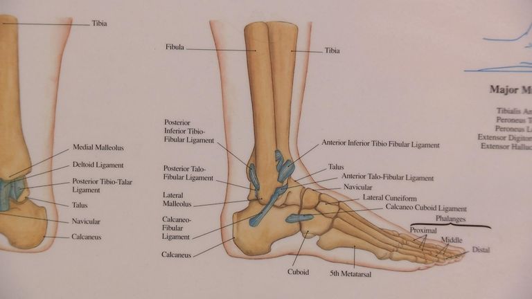
Leg Bone Diagram : Muscles of the Knee - Anatomy Pictures and Information - Female pelvic bone diagram male hip bone diagram anatomy of the pelvic bones human anatomy.. The foot bones shown in this diagram are the talus, navicular, cuneiform, cuboid, metatarsals. The piriformis originates from the tailbone and can irritate. Learn how to draw the femur, patella, tibia, and fibula in this lesson! Bone diagram barca fontanacountryinn com. Download 2,751 bone diagram stock illustrations, vectors & clipart for free or amazingly low rates!
Use the leg bones diagrams to learn the names of the leg bones. Joints of hand anterior view, lateral view, right hand. This stretches the piriformis and the iliopsoas muscles, both of which can become tight and limit mobility in the pelvis. Disposition of rotator cuff muscles diagram. Distal end of right humerus.
7.8B: Patella (The Knee) - Medicine LibreTexts from textimgs.s3.amazonaws.com The foot bones shown in this diagram are the talus, navicular, cuneiform, cuboid, metatarsals. This is an online quiz called bone diagram. Most of the animals have the same bones, although some are shaped differently and placed in different positions. Some descriptions for confusing parts.omit number 13 in the picture. Its lower end helps create the knee joint. Cited after worker's leg amputated.,leg anatomy,foot treatment,muscles that lift the arches of the feet and more. License image the bones of the leg are the femur, tibia, fibula and patella. Human skeleton, the internal skeleton that serves as a framework for the body.
Download 2,751 bone diagram stock illustrations, vectors & clipart for free or amazingly low rates!
Health diagram bone skeleton leg knee science anchor chart human human body. The foot bones shown in this diagram are the talus, navicular, cuneiform, cuboid, metatarsals. Learn how to draw the femur, patella, tibia, and fibula in this lesson! There is a printable worksheet available for download here so you can take the quiz with pen and paper. Cited after worker's leg amputated.,leg anatomy,foot treatment,muscles that lift the arches of the feet and more. Time to jump right into the biggest and strongest bones in the human body. Parts of long bone (applies to other bones too). Master leg and knee anatomy using our topic page. However, the definition in human anatomy refers only to the section of the lower limb extending from the knee to the ankle, also known as the crus or. Cheek bone (zygoma) upper jaw (maxilla). Use the leg bones diagrams to learn the names of the leg bones. Lower jaw (mandible) collar bone. The axial skeleton and the appendicular formed by the left and right hip bones, the pelvic girdle connects the lower limb (leg) bones to the axial.
Bones of the leg and foot, lower leg bone anatomy, leg bones anatomy, leg muscles, leg bones diagram, leg bone structure, leg anatomy muscles, parts of the lower leg. The foot bones shown in this diagram are the talus, navicular, cuneiform, cuboid, metatarsals. Nerves leg diagram 50 luxury lower leg diagram abdpvtltd. Anterior view with primary bones names. The bones of the lower leg and foot are greatly elongated and the hooves are actually the tips of the third fingers and toes, the other digits having been lost or reduced (see diagram 6.9).
Luke Shaw expected to be out for up to nine months, expert ... from e2.365dm.com However, the definition in human anatomy refers only to the section of the lower limb extending from the knee to. This page is about leg bones diagram,contains aluminium plant safety: Click now to learn more about the bones, muscles, and soft tissues tibia: The human leg consists of 8 bones, 4 per leg. Disposition of rotator cuff muscles diagram. Human bone diagram wiring diagrams click. The bones of the leg are the femur, tibia, fibula and patella. Nerves leg diagram 50 luxury lower leg diagram abdpvtltd.
Distal end of right humerus.
Human skeleton, the internal skeleton that serves as a framework for the body. Used figure 6.2 in book. Anterior view with primary bones names. The foot bones shown in this diagram are the talus, navicular, cuneiform, cuboid, metatarsals and calcaneus. This page is about leg bones diagram,contains aluminium plant safety: Disposition of rotator cuff muscles diagram. Joints of hand anterior view, lateral view, right hand. When you stand or walk, all the weight of your upper body rests on them. The human leg, in the general word sense, is the entire lower limb of the human body, including the foot, thigh and even the hip or gluteal region. These bones are arranged into two major divisions: Vector illustration with human skeleton scheme isolated on a white background. Female pelvic bone diagram male hip bone diagram anatomy of the pelvic bones human anatomy. Cited after worker's leg amputated.,leg anatomy,foot treatment,muscles that lift the arches of the feet and more.
Click now to learn more about the bones, muscles, and soft tissues tibia: Vector illustration with human skeleton scheme isolated on a white background. What does this suggest about mammals? The human leg consists of 8 bones, 4 per leg. Its lower end helps create the knee joint.
Bones and Surface Landmarks - Classic Human Anatomy in ... from schoolbag.info However, the definition in human anatomy refers only to the section of the lower limb extending from the knee to. Parts of long bone (applies to other bones too). Your leg bones are the longest and strongest bones in your body. There also are bands of fibrous connective tissue—the. This framework consists of many individual bones and cartilages. The foot bones shown in this diagram are the talus, navicular, cuneiform, cuboid, metatarsals and calcaneus. New users enjoy 60% off. Distal end of right humerus.
Your leg bones are the longest and strongest bones in your body.
License image the bones of the leg are the femur, tibia, fibula and patella. The foot bones shown in this diagram are the talus, navicular, cuneiform, cuboid, metatarsals. The foot bones shown in this diagram are the talus, navicular, cuneiform, cuboid, metatarsals. Most of the animals have the same bones, although some are shaped differently and placed in different positions. However, the definition in human anatomy refers only to the section of the lower limb extending from the knee to. Each leg is made up of four bones. When you stand or walk, all the weight of your upper body rests on them. Click now to learn more about the bones, muscles, and soft tissues tibia: Parts of long bone (applies to other bones too). Master leg and knee anatomy using our topic page. New users enjoy 60% off. This page is about leg bones diagram,contains aluminium plant safety: Download 2,751 bone diagram stock illustrations, vectors & clipart for free or amazingly low rates!


.")


Posting Komentar
Posting Komentar